


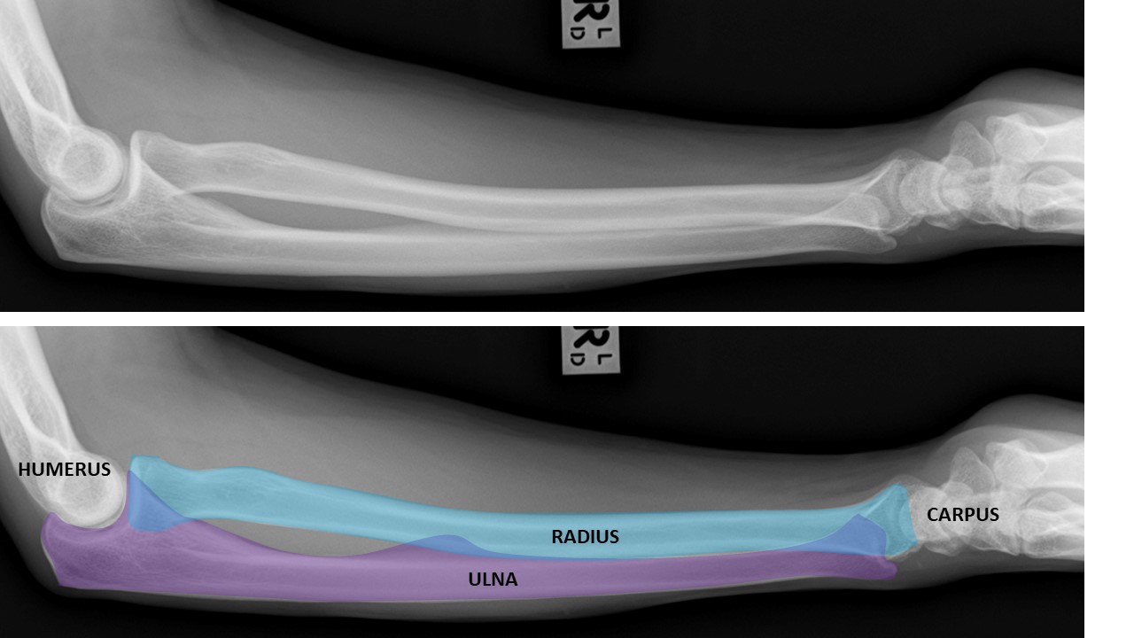
The FOREARM is composed of the RADIUS and the ULNA. They are paired long bones.
The radius and ulna articulate with the HUMERUS proximally and the CARPUS distally.
This is an AP projection of the forearm. [Courtesy of Jessica Hui Shi Ng, Radiopaedia.org, rID: 73169]

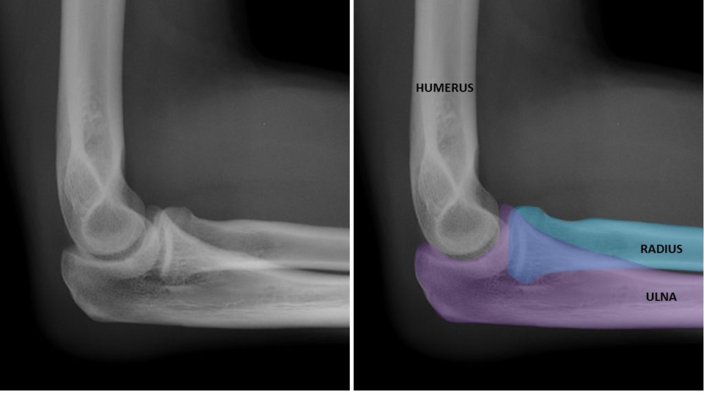
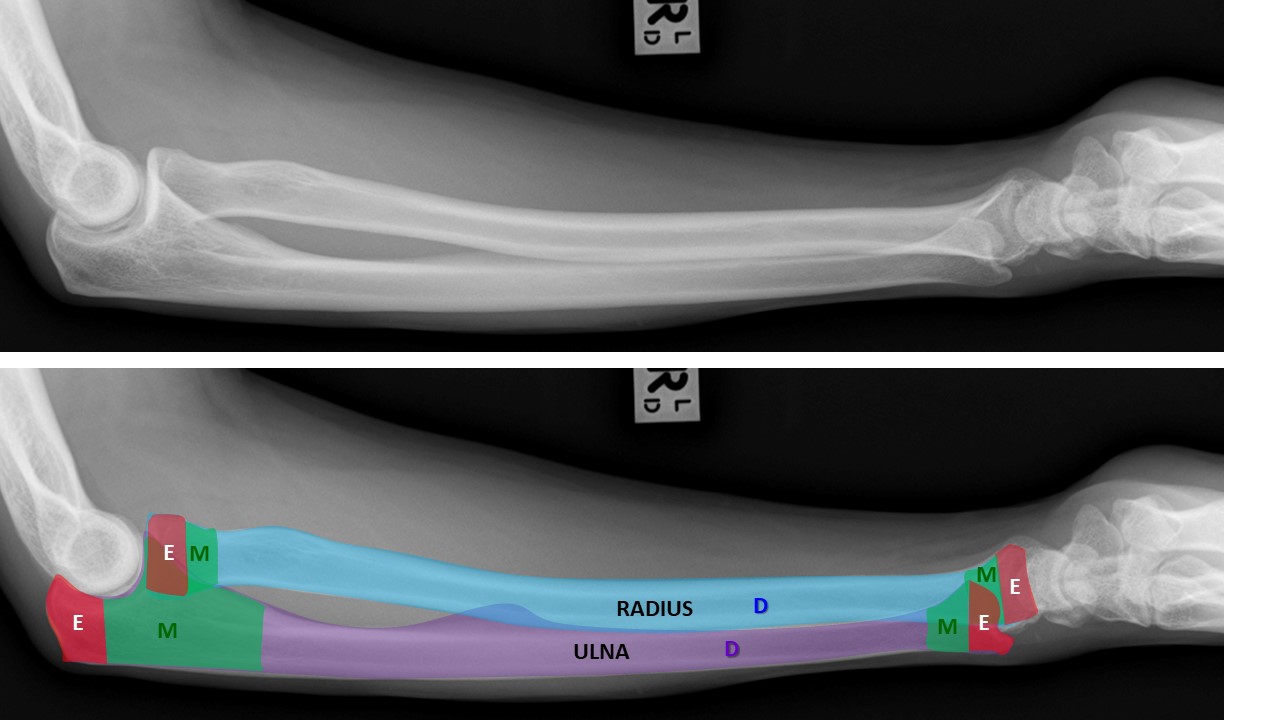
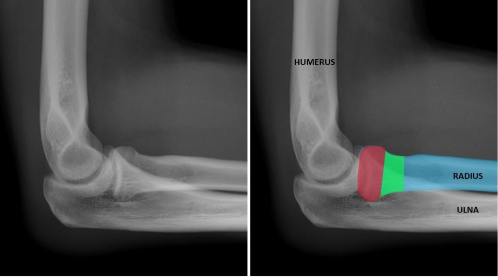
This is a Lateral projection of the forearm. [Courtesy of Jessica Hui Shi Ng, Radiopaedia.org, rID: 73169]
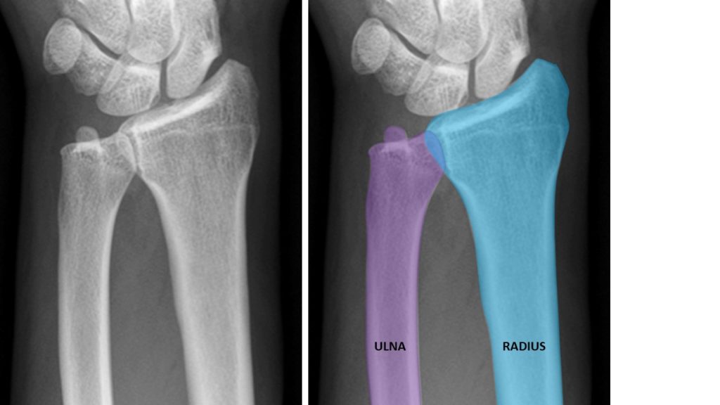
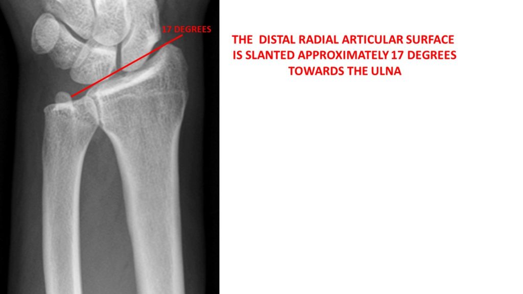
AP view distally as it articulates with the wrist.
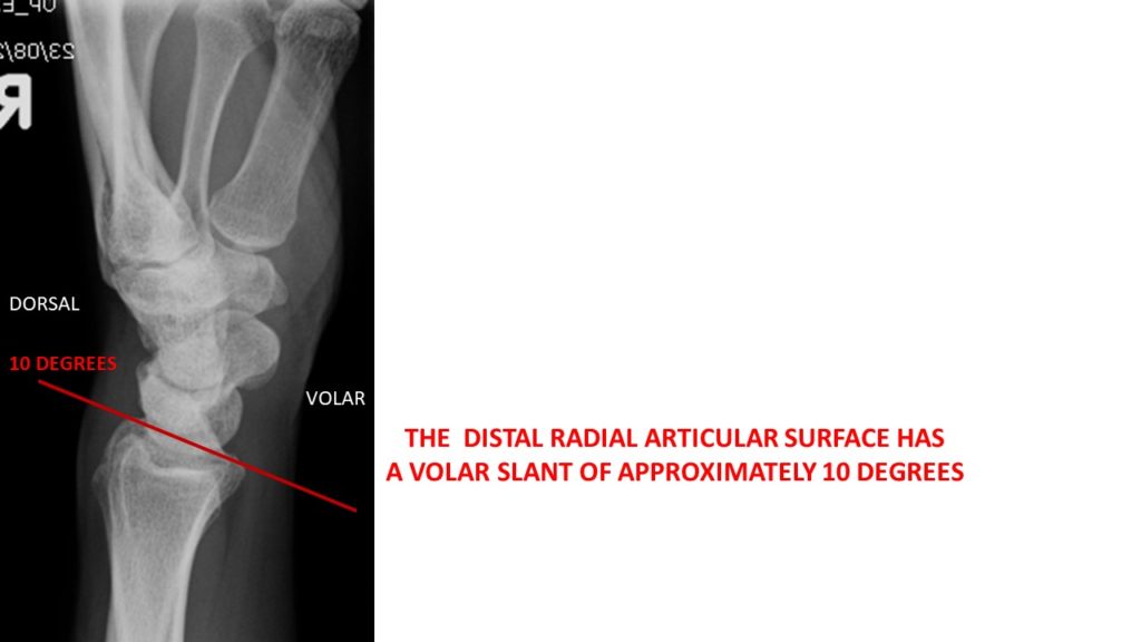
Lateral view distally as it articulates with the wrist.
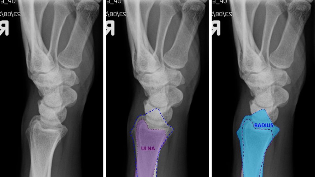
The RADIUS (BLUE & BLUE DASHES) and ULNA (PURPLE & PURPLE DASHES) overlap (superimposed) upon one another in the lateral projection.
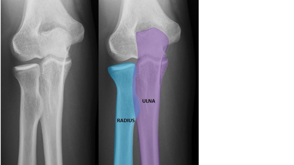
AP view proximally as it articulates with the humerus.
Lateral view proximally as it articulates with the humerus.
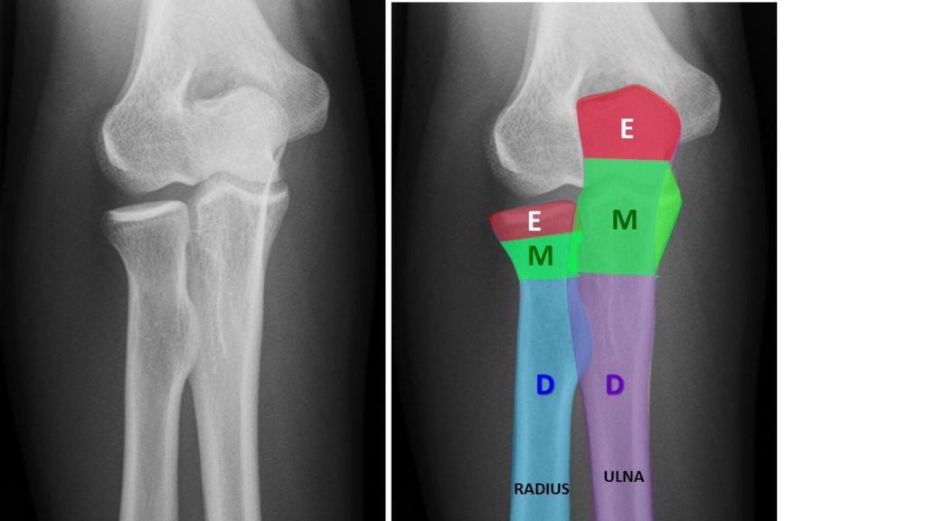
The RADIUS and the ULNA are classified as LONG BONES.
The RADIUS and ULNA each have
- EPIPHYSIS (proximal and distal)
- METAPHYSIS (proximal and distal) and
- DIAPHYSIS (aka SHAFT)
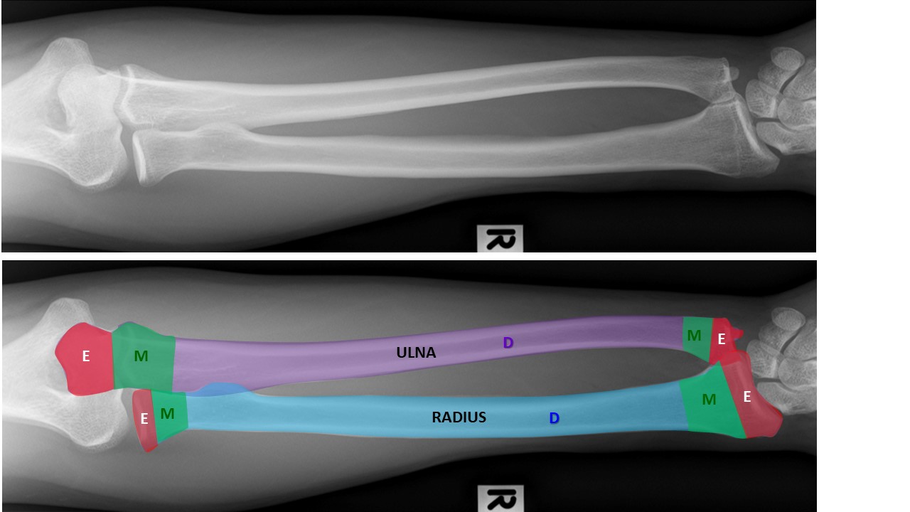
EPIPHYSIS: at the ends of the long bones (E)
METAPHYSIS: the fluted portions between the epiphyses and shaft (M)
DIAPHYSIS: the shaft of the long bone (D) (D)
Distal forearm
- depicting distal epiphyses, distal metaphyses, and distal diaphyses. AP view

Lateral view

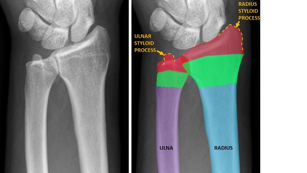
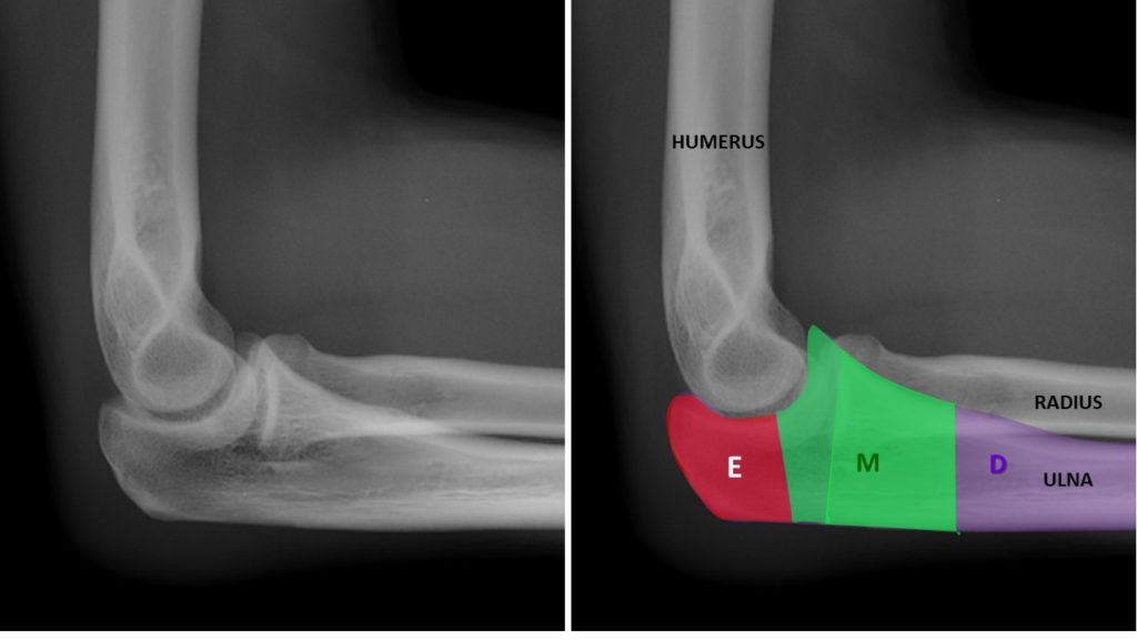
Distal end of forearm
The distal end of the forearm demonstrates the following anatomy:
RADIUS:
- distal epiphysis (RED) with RADIAL STYLOID PROCESS (YELLOW DOTTED LINE)
- distal metaphysis (GREEN)
- distal diaphysis (BLUE)
ULNA:
- distal epiphysis aka ULNAR HEAD (RED) with ULNAR STYLOID PROCESS (YELLOW DOTTED LINE)
- distal metaphysis aka ULNAR NECK (GREEN)
- distal diaphysis (PURPLE)
AP view
Lateral view
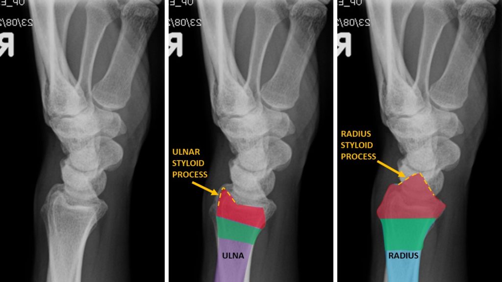
NOTE: The distal radius is much larger than the distal ulna.
SIDE NOTE: There are multiple STYLOID PROCESSES in the human skeleton. A styloid process is a slender pointed piece of bone. (from Greek stylos (στῦλος), “pillar”).
Proximal forearm
- depicting proximal epiphyses, proximal metaphyses, and proximal diaphyses.
AP view
Lateral view

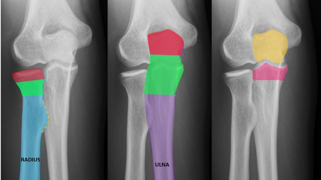
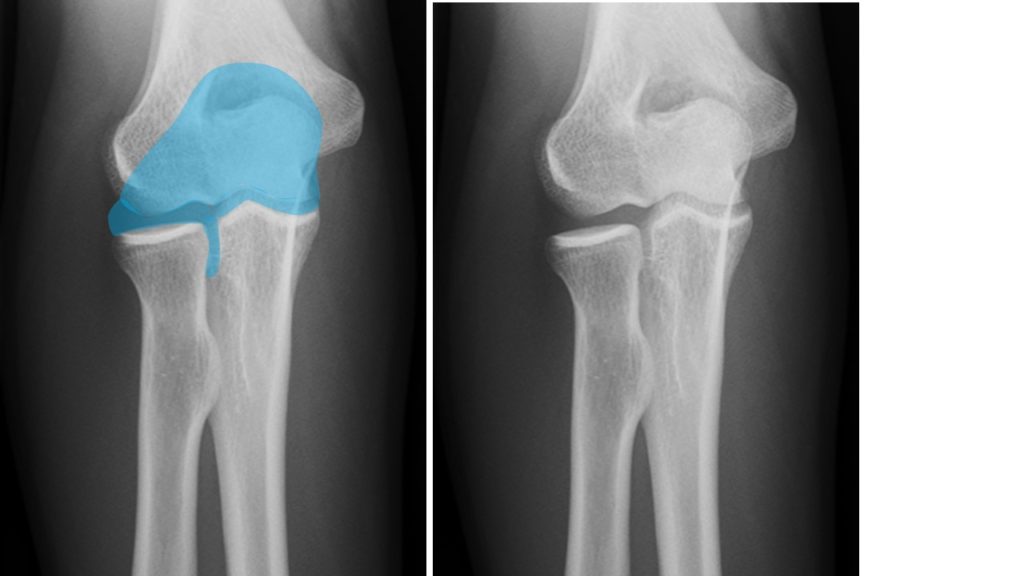
Proximal end of forearm
The proximal end of the forearm demonstrates the following anatomy:
RADIUS:
- proximal epiphysis aka RADIAL HEAD (RED) which articulates with the capitellum of humerus
- proximal metaphysis aka RADIAL NECK (GREEN)
- proximal diaphysis (BLUE)
- RADIAL TUBEROSITY, (YELLOW DOTTED LINE) an eminence of the medial aspect; posterior portion is for the attachment of biceps brachii; anterior portion on which a bursa is interposed between the tendon and the bone.
ULNAR:
- proximal epiphysis (RED) which articulates with the olecranon fossa of humerus
- proximal metaphysis (GREEN)
- proximal diaphysis (PURPLE)
- OLECRANON PROCESS (YELLOW) which articulates with the olecranon fossa of humerus
- CORONOID PROCESS (PINK) which articulates with the trochlea of humerus
AP view
Lateral view
NOTE: The proximal ulna is much larger than the proximal radius.

- OLECRANON PROCESS (YELLOW) which articulates with the olecranon fossa of humerus
- CORONOID PROCESS (PINK) which articulates with the trochlea of humerus

OSSEOUS RELATIONSHIPS
RELATIONSHIP BETWEEN HUMERUS AND RADIUS
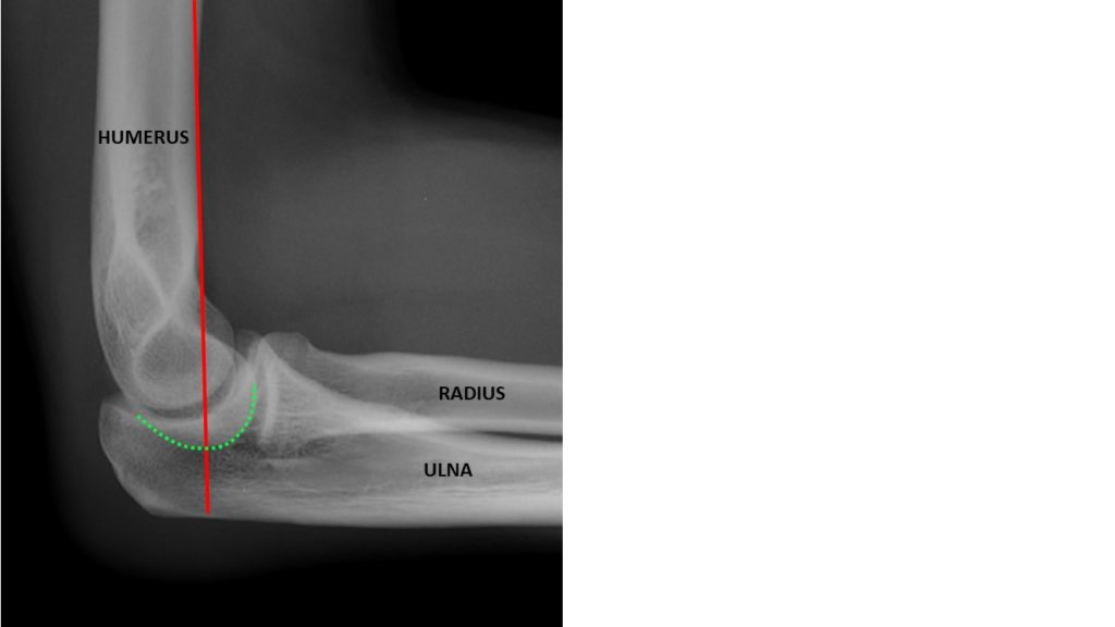
RADIOCAPITELLAR LINE:
A line drawn through the CENTER OF THE RADIAL NECK should pass through the CENTER OF THE CAPITELLUM, whatever the positioning of the patient, since the radius articulates with the capitellum.
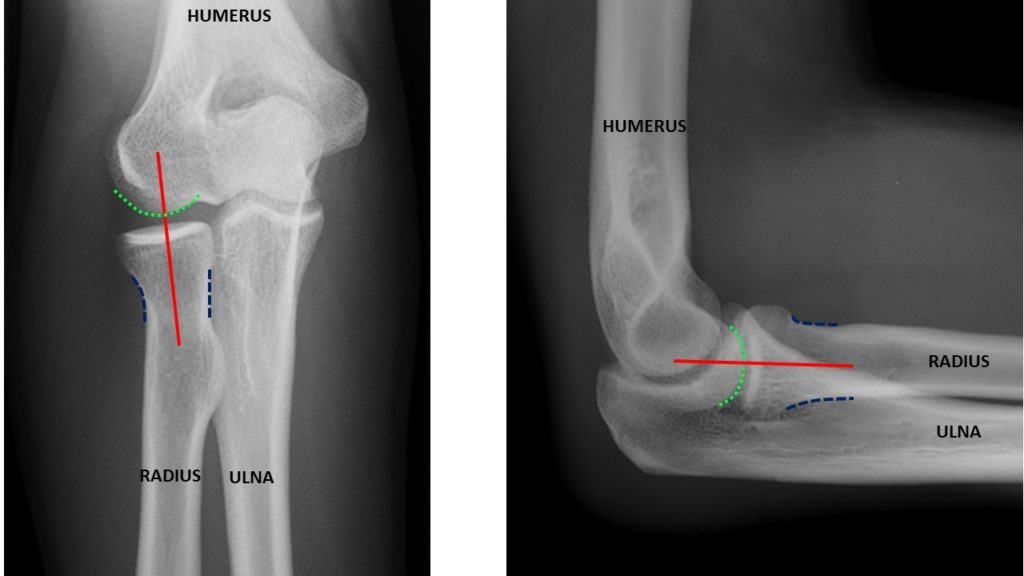
ANTERIOR HUMERAL LINE:
On the lateral elbow projection, a line constructed immediately anterior to humerus should pass through the MIDDLE THIRD OF THE CAPITELLUM.
CORTEX: External layer of compact lamellar bone
- COMPOSITION – The OSTEON is the functional unit of compact bone, and contain LAMELLAE, CENTRAL CANAL, LACUNAE, and CANALICULI.
MEDULLARY: Internal trabecular / spongy / cancellous bone
- COMPOSITION – Mineralized bars of osseous tissue with numerous spaces in extracellular matrix that contain red bone marrow.


ARTHROLOGY / WRIST
The DISTAL RADIOULNAR ARTICULATION is a SYNOVIAL PIVOT-TYPE JOINT between the two bones in the forearm; the radius and ulna.
The RADIOCARPAL JOINT is a CONDYLOID ARTICULATION.
The ARTICULAR DISK (aka TRIANGULAR FIBROCARTILAGE) is triangular in shape, and is placed transversely distal to the head of the ulna.
- Its proximal surface articulates with the head of the ulna, forming an ARTHRODIAL JOINT;
- Its distal surface articulates with the triquetrum and medial part of the lunate.

ARTHROLOGY / ELBOW
The elbow articulation is a compound SYNOVIAL joint classsified as a HINGE JOINT.
There are three joints in the elbow.
- The HUMERORADIAL JOINT: articulation between the CAPITELLUM of distal humerus and HEAD of proximal radius.
- The HUMEROULNAR JOINT: articulation between the TROCHLEA of distal humerus and TROCHLEAR NOTCH on proximal ulna.
- The PROXIMAL RADIOULNAR JOINT: articulation between the HEAD of proximal radius and the RADIAL NOTCH of proximal ulna.

Coming up… the HUMERUS!