


Now onto the wrist! The wrist is composed of 8 carpal bones.
Each carpal bone, with the exception of pisiform, presents 6 surfaces.
The structure of the carpal bones is cancellous tissue enclosed in a layer of compact bone.
There are 2 rows of carpal bones: the DISTAL and PROXIMAL rows. Each row has 4 carpal bones.
When radiographs are taken of the wrist, at least 3 views are needed for complete and accurate interpretation.
These views are: PA (posterior to anterior); Medial Oblique; and Lateral.
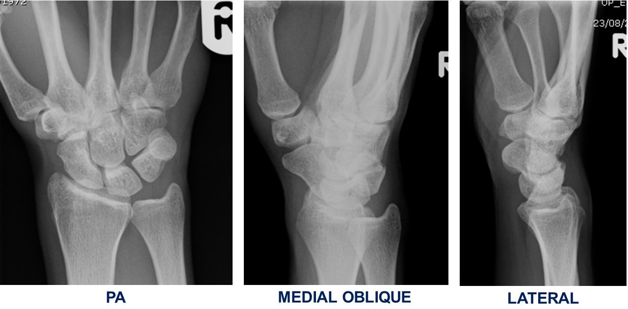
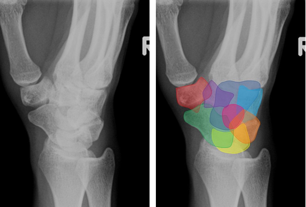
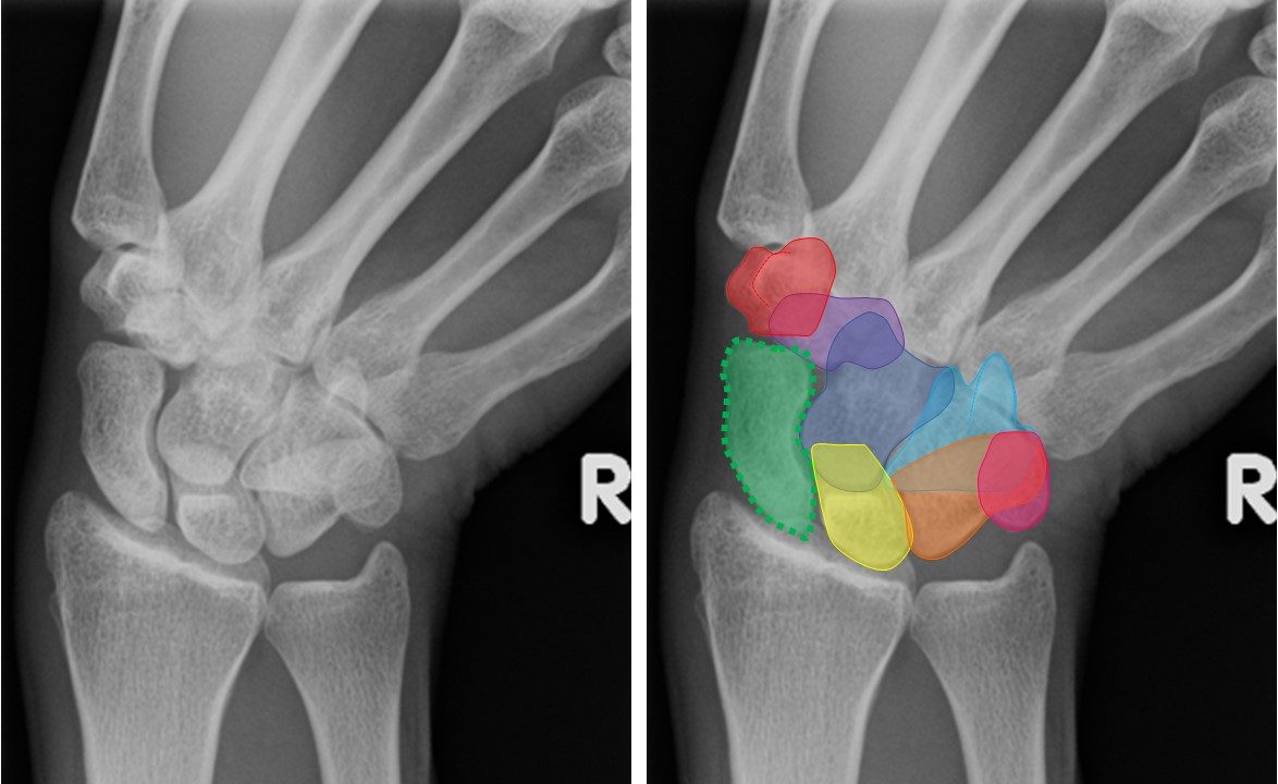
The view below is a PA view without and with colored/outlined carpal bones.
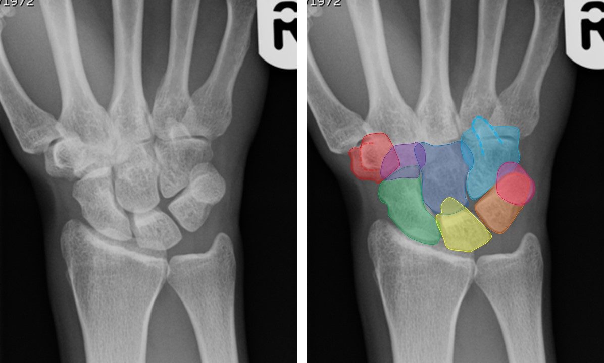
The DISTAL CARPAL ROW is composed of:
TRAPEZIUM (aka GREATER MULTANGULAR) RED
TRAPEZOID (aka LESSER MULTANGULAR) PURPLE
CAPITATE DARK BLUE
HAMATE LIGHT BLUE
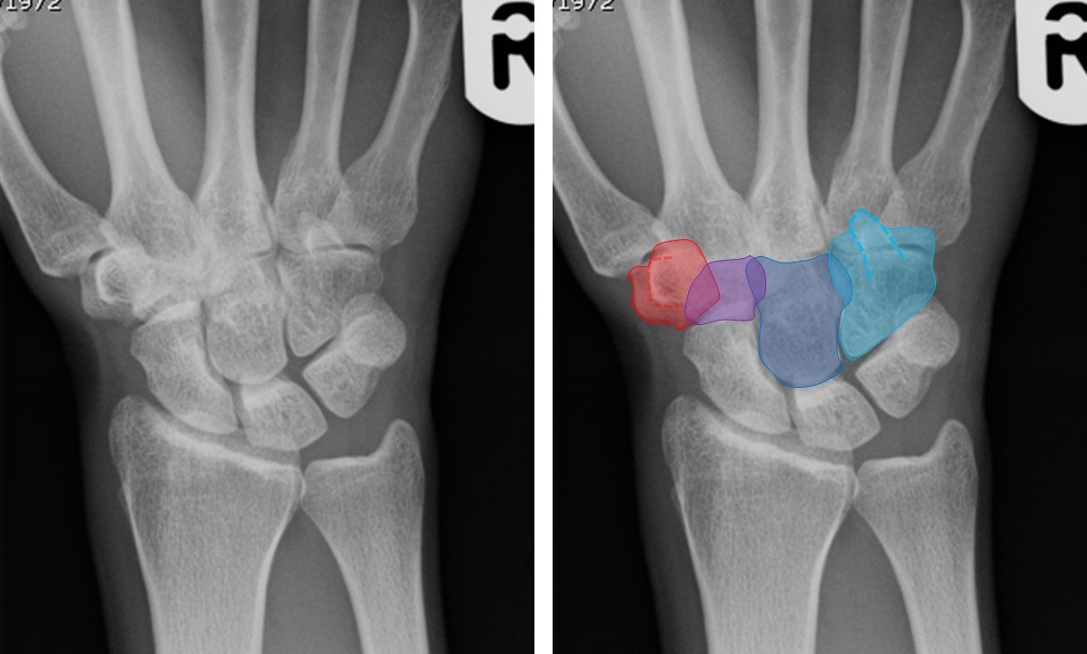
The PROXIMAL ROW is composed of:
SCAPHOID (aka NAVICULAR) GREEN
LUNATE YELLOW
TRIQUETRUM (aka TRIANGULAR) ORANGE
PISIFORM PINK
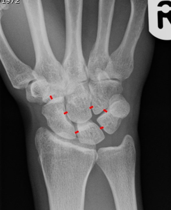
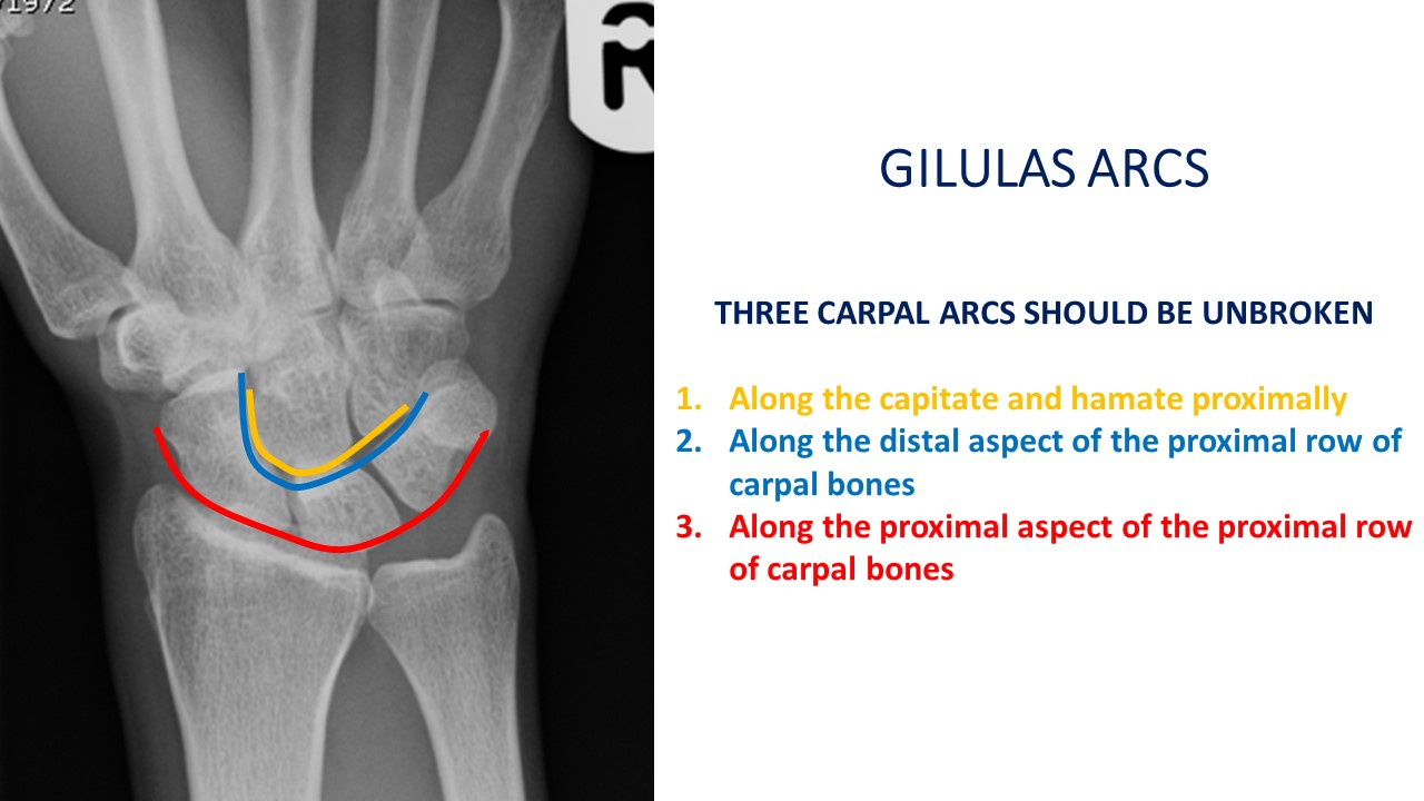
NORMAL ANATOMICAL RELATIONSHIPS
The width between the carpal bones are uniform and approximately 2 mm (paediatric wrists exceptions).
The distal radial articular surface is slanted approximately 17 degrees towards the ulna.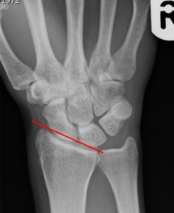
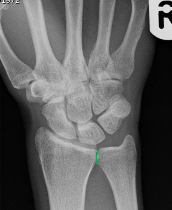
RADIOULNAR JOINT
Medially the distal radius articulates with the head of ulna at the ulnar notch.
The head of ulna is usually 2 mm shorter than the radius and eithers touches or slightly overlaps the radius at the distal radioulnar joint.
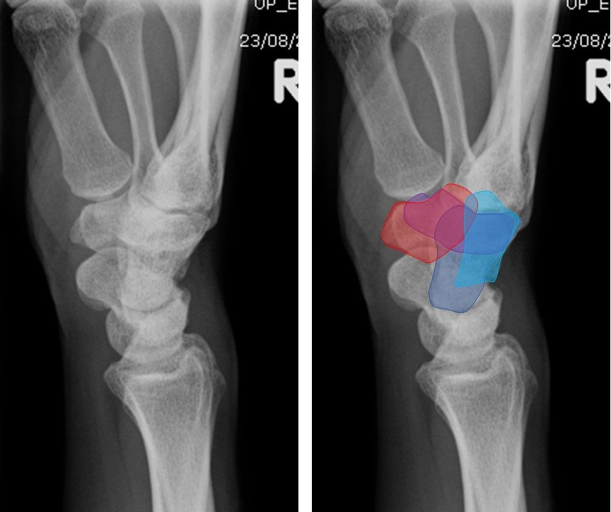
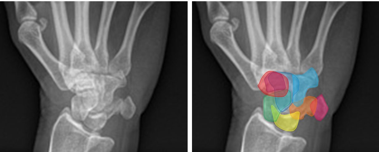
The view below is a Medial Oblique view without and with colored/outlined carpal bones.
The DISTAL CARPAL ROW is composed of:
TRAPEZIUM (aka GREATER MULTANGULAR) RED
TRAPEZOID (aka LESSER MULTANGULAR) PURPLE
CAPITATE DARK BLUE
HAMATE LIGHT BLUE
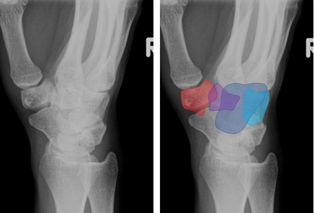
The PROXIMAL ROW is composed of:
SCAPHOID (aka NAVICULAR) GREEN
LUNATE YELLOW
TRIQUETRUM (aka TRIANGULAR) ORANGE
PISIFORM PINK
The TRAPEZIUM and DISTAL SCAPHOID are well visualized in the medial oblique proection.
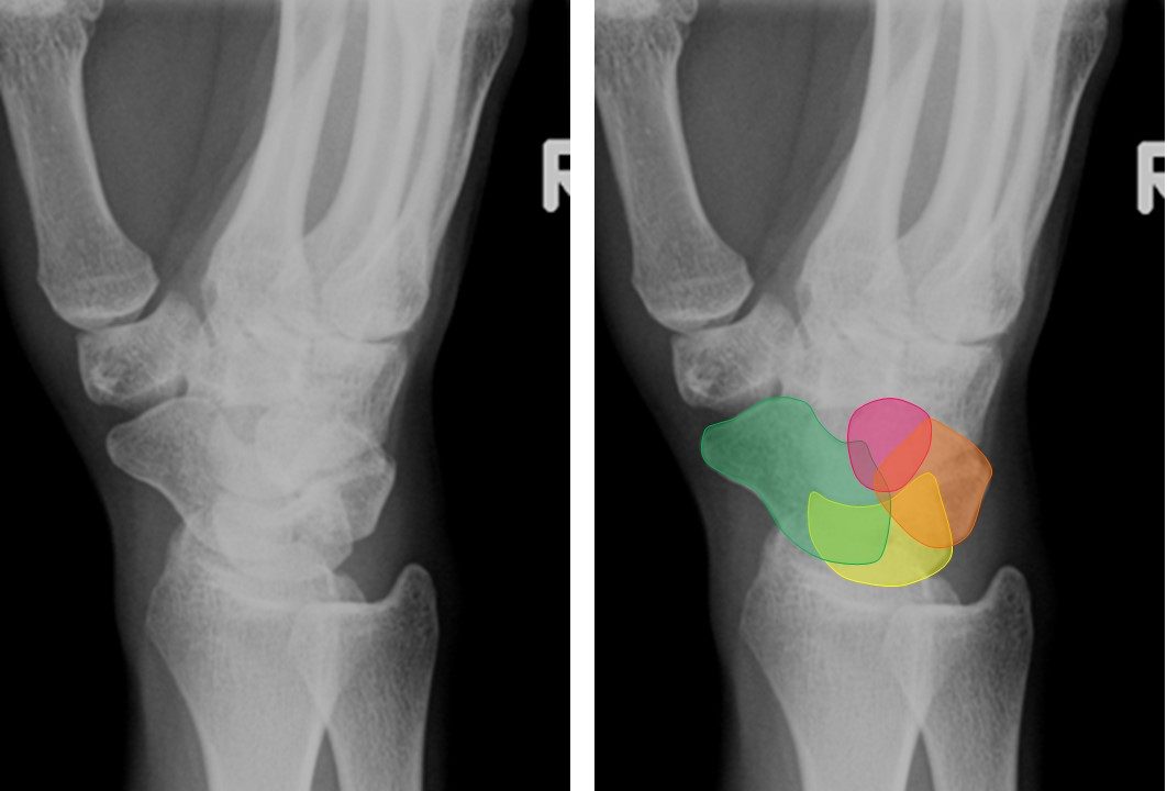
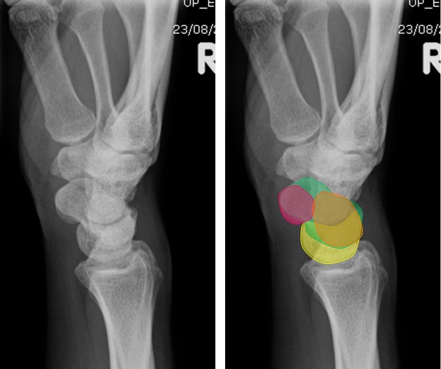
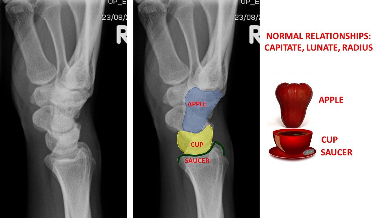
The view below is a Lateral view without and with colored/outlined carpal bones.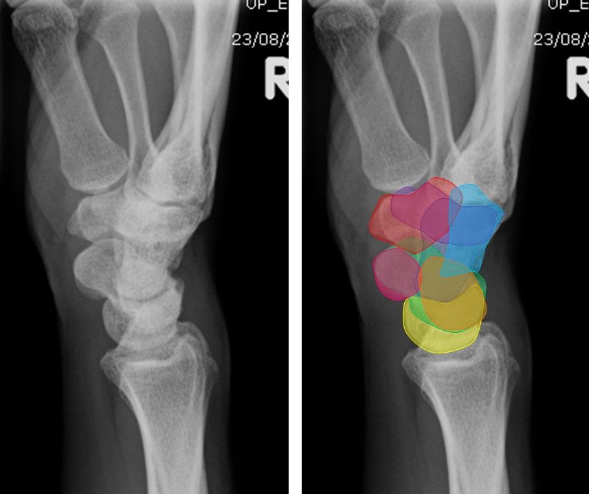
The DISTAL CARPAL ROW is composed of:
TRAPEZIUM (aka GREATER MULTANGULAR) RED
TRAPEZOID (aka LESSER MULTANGULAR) PURPLE
CAPITATE DARK BLUE
HAMATE LIGHT BLUE
The PROXIMAL ROW is composed of:
SCAPHOID (aka NAVICULAR) GREEN
LUNATE YELLOW
TRIQUETRUM (aka TRIANGULAR) ORANGE
PISIFORM PINK
NORMAL ANATOMICAL RELATIONSHIPS
On the Lateral projection the CAPITATE, LUNATE, and RADIAL SURFACE should line up.
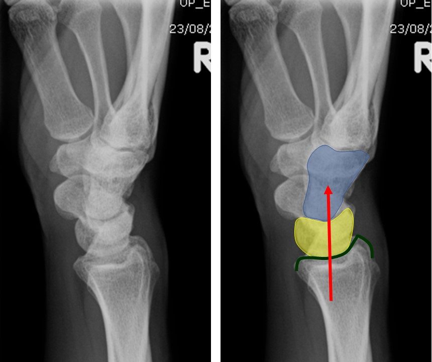
To easily remember this relationship the CAPITATE has been likened to the thai rose apple that sits in a cup (the LUNATE) which sits on top of a saucer (the RADIAL SURFACE).
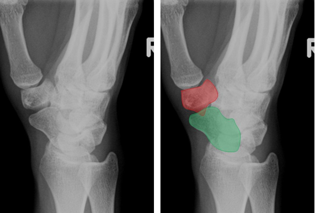
VOLAR TILT

At times additional x-rays maybe required to better assess certain anatomical structures. Two of these views are the PA view with Ulnar Flexion and a Lateral Oblique.
PA ULNAR FLEXION: this is the best view to visualize the SCAPHOID.
POSTERIOR OBLIQUE (aka BALL CATCHER’S VIEW; NORGAARD’S VIEW): Typically taken bilaterally.
This is the best view to visualize the PISIFORM.
INTERESTING ANATOMICAL FACTS OF EACH CARPAL BONE:
TRAPEZIUM: 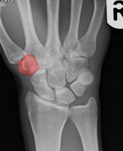
The trapezium is also know as GREATER MULTANGULAR.
- The trapezium articulates with four bones:
- the navicular
- the first metacarpal
- the trapezoid (aka lesser multangular)
- the second metacarpal
=====
TRAPEZOID: 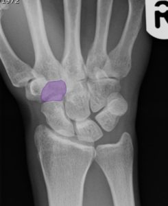
The trapezoid is also known as the LESSER MULTANGULAR.
The trapezoid is the smallest bone in the distal carpal row.
- The trapezoid articulates with four bones:
- the navicular
- the second metacarpal
- the trapezium (aka greater multangular)
- the capitate
=====
CAPITATE: 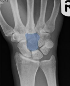
The capitate is the largest of the carpal bones and occupies the center of the wrist.
- The capitate articulates with seven bones:
- the navicular
- the lunate
- the second metacarpal
- the third metacarpal
- the fourth metacarpal
- the trapezoid
- the hamate
=====
HAMATE: 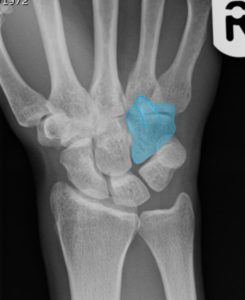
The hamate bone is notable by its wedge-shaped form and the hook-like process, known as the HAMULUS, which projects from its volar surface.
- The hamate articulates with five bones:
- the lunate
- the fourth metacarpal
- the fifth metacarpal
- the triquetrum
- the capitate
=====
SCAPHOID: 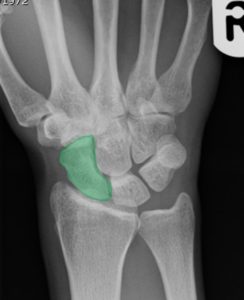
The scaphoid is also known as NAVICULAR.
The scaphoid bone is the largest bone of the proximal row.
It received its name from its resemblance to a boat.
- The scaphoid articulates with five bones:
- the radius
- the trapezium
- the trapezoid
- the capitate
- the lunate
=====
LUNATE: 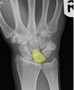
The lunate bone may be distinguished by its deep concavity and crescentic outline.
- The lunate articulates with five bones:
- the radius
- the capitate
- the hamate
- the scaphoid
- the triquetrum
=====
TRIQUETRUM: 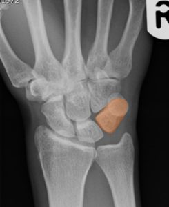
The triquetrum is also known as TRIANGULAR bone.
The triquetrum bone is apparent by its pyramidal shape and by an oval isolated facet for articulation with the pisiform bone.
- The triquetrum articulates with three bones:
- the lunate
- the pisiform
- the hamate
- plus – the triangular articular disk which separates it from the lower end of the ulna
=====
PISIFORM: 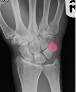
The pisiform bone is known by its small size and by its single articular facet.
- The pisiform articulates with one bone:
- the triquetrum
=====
ARTICULATIONS:
Carpal articulations may be subdivided into three sets:
- The articulations of the Proximal Row of Carpal Bones
- The wrist joint also referred to as the radiocarpal joint is a CONDYLOID SYNOVIAL JOINT.
- The articulations of the Distal Row of Carpal Bones
- The carpometacarpal joints (CMC) are SYNOVIAL PLANE JOINTS.
- The carpometacarpal of the thumb is a SADDLE JOINT.
- The articulations of the Two Rows with each Other
- The intercarpal joints are the SYNOVIAL PLANE JOINTS.
Synovial Membrane.—The synovial membrane of the carpus is very extensive, and bounds a synovial cavity of very irregular shape.